Filtered by author: Elailne Song Clear Filter

The Promise and Responsibility of Artificial Intelligence in Wound Care

Introduction

Advancements in artificial intelligence (AI) are happening rapidly across healthcare, and wound care is no exception. While AI-driven technologies are not here to replace clinicians, they have the potential to be powerful tools - enhancing decision-making, improving efficiencies, and ultimately leading to better patient outcomes.

As with any new development in healthcare, the key to successful integration lies in a balanced approach: embracing innovation while ensuring safety, reliability, and responsible implementation. In this blog, we provide a brief overview of the evolution of AI in healthcare, its current and future applications in wound care, and how clinicians can best navigate this technological shift.

For details, please refer to the Webinar “The Promise and Responsibility of AI in Wound Care”, available for free to all AAWC Members.

The Origins of Artificial Intelligence in Healthcare

Artificial intelligence, as a concept, dates back to the mid-20th century. Early AI models were built on the idea of mimicking human intelligence through algorithms and computational learning. Over time, AI progressed from simple rule-based systems to more sophisticated machine learning and deep learning models, capable of analyzing complex datasets, recognizing patterns, and making predictive recommendations.

In healthcare, AI's journey began with clinical decision support systems and diagnostic algorithms, but recent advancements have expanded its role significantly - particularly in areas such as imaging, diagnostics, and personalized medicine.

The Evolution of AI in Healthcare

The term "Artificial Intelligence" was coined by John McCarthy in 1955, a computer science professor at Stanford University. Since then, AI has evolved a lot and the development of AI in healthcare has progressed through three key epochs:

 AI 1.0 – Symbolic and Probabilistic Models

This early phase focused on rule-based systems, where AI was programmed with predefined rules to assist in clinical decision-making. While useful, these systems were limited in their ability to adapt or learn from new data. Examples include traditional ‘expert system’-based clinical decision support tools.

AI 2.0 – Machine Learning and Deep Learning Models

The introduction of machine learning enabled AI systems to learn from data, improving performance over time. With classification, prediction, and pattern recognition capabilities, this era saw the rise of sophisticated diagnostic tools, predictive analytics, and personalized treatment plans. Examples include AI models that help classify diabetic foot ulcers.

AI 3.0 – Generative AI

Today, we are in the third epoch of AI, characterized by generative AI, which can create human-like text, images, and even treatment plans. While highly flexible and powerful, generative AI also comes with risks, such as hallucinations (inaccurate or made-up information). Ensuring clinical judgment and rigorous validation are imperative for AI integration in clinical practice. Examples include PubMedGPT.

Current Applications of AI in Wound Care

Wound care can be categorized into wound diagnosis, wound management, and wound prognosis & prevention. In the webinar, we introduce a framework that highlights the potential benefits of AI-driven technologies from epochs 2.0 and 3.0 in these key areas.

Refer to the Webinar “The Promise and Responsibility of AI in Wound Care” to explore how AI is being leveraged to:

  • Improve clinical decision-making by providing clinicians with faster and more accurate access to critical information.
  • Predict healing outcomes with advanced machine learning models.
  • Support clinicians in optimizing wound diagnosis, prognosis, and management.

Ensuring Responsible AI Use in Wound Care

While AI offers many benefits, it is crucial to acknowledge its limitations. The potential for AI to generate incorrect or misleading information—known as AI hallucinations—poses a significant risk in healthcare.

Responsible AI use requires a robust framework that ensures AI tools are accurate, reliable, and transparent. It involves:

  • Rigorous Testing & Validation – Ensuring AI tools are accurate, reliable, and transparent.
  • Continuous Monitoring – Regular assessment of AI performance to maintain reliability.
  • Clinician Education – Training healthcare professionals on AI’s limitations and its role as a support tool rather than a replacement for human judgment.

In the webinar, we also share our journey in developing an AI-powered clinical intelligence solution to help clinicians quickly access trusted, evidence-based wound care information. The resulting AI model is specifically trained to prevent hallucinations, and ensure that every answer comes directly from a carefully vetted knowledge base. This knowledge base is built through a rigorous, unbiased editorial process, guaranteeing both accuracy and reliability.

Key Considerations for Responsible AI in Wound Care

During the webinar, we also discussed critical considerations for AI adoption in wound care, including:

  • Data Collection & Accessibility – Ensuring diverse and representative datasets.
  • Algorithm Training & Diversity – Preventing bias in AI models.
  • Ethical & Legal Concerns – Addressing liability and regulatory challenges.
  • Data Privacy & Security – Safeguarding sensitive patient information.
  • Equitable Access & Cost – Ensuring AI tools remain accessible to all healthcare clinicians.

Conclusion

The integration of AI into wound care is promising, but it requires a balanced approach. Staying open to the advancements AI can bring, while proactively addressing challenges, is critical to ensuring the safe and effective use of these technologies.

It is also essential to remember that these tools are meant to support, not replace, human judgment. Moreover, AI should be used to enhance, not undermine, the patient-clinician relationship. In wound care, where trust and communication are critical, it is essential that AI tools are used in a way that supports, rather than disrupts, this relationship.

References

  • Chen M-Y, Cao M-Q, Xu T-Y. Progress in the application of artificial intelligence in skin wound assessment and prediction of healing time. Am J Transl Res. 2024 Jul 15;16(7):2765–76
  • Ganesan O, Morris MX, Guo L, Orgill D. A review of artificial intelligence in wound care. Artif Intell Surg. 2024 Nov 4;4(4):364–75.
  • Howell MD, Corrado GS, DeSalvo KB. Three epochs of artificial intelligence in health care. JAMA. 2024 Jan 16;331(3):242–4.
  • Rippon MG, Fleming L, Chen T, Rogers AA, Ousey K. Artificial intelligence in wound care: diagnosis, assessment and treatment of hard-to-heal wounds: a narrative review. J Wound Care. 2024 Apr 2;33(4):229–42.
  • Song EH, Milne C, Lucchese AC. The Promise and Responsibility of AI in Wound Care: Enhancing Clinical Decision-Making. Accessed on 2/21/25 from https://woundreference.com/blog?id=responsible-ai

Patient Case Study: Jose Doe's Journey With Squamous Cell Carcinoma In Situ

Background

At the DeLand Foot and Leg Center, we treat patients with complex medical histories, such as Jose Doe. His journey is particularly challenging as it involves chronic conditions like diabetes, a below-knee amputation, and the fight against squamous cell carcinoma in situ. Jose's case is a prime example of the intricate relationship between multiple health issues and underscores the importance of tailoring treatment plans to each individual's unique circumstances.

Patient Presentation

Jose, a 67-year-old male, came to us with a significant medical history consisting of diabetes, peripheral vascular disease, and an above-knee amputation. He presented with a plantar lesion that had not improved despite being previously treated as a wart by another care provider. Given the lesion's persistence for over three months and lack of response to treatment, a biopsy was performed during his initial visit. His condition is further complicated by chronic arterial insufficiency that necessitates stents and obesity.

Clinical Observations

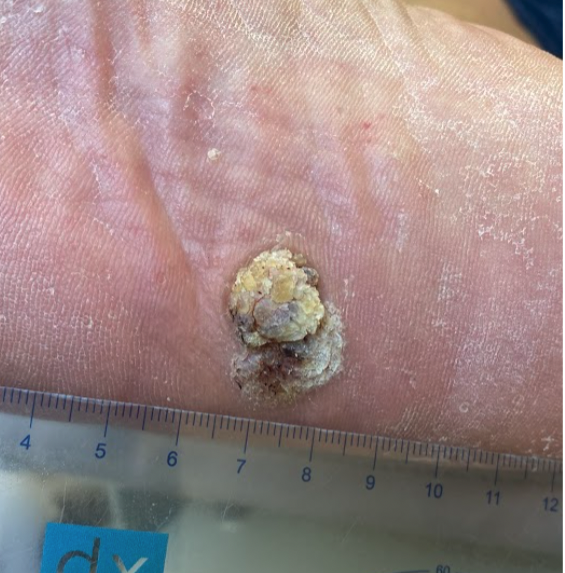
Shave biopsies of the right foot's lesion, measuring 2.1 cm wide x 1.4 cm long x 0.3 cm elevated, revealed a diagnosis of squamous cell carcinoma in situ (Bowen's Disease). The location of the lesion on the midfoot plantar surface with an expanding margin on the lateral side is troublesome because this is the patient's right foot, which is his only weight-bearing foot as a result of the above-knee amputation (AKA). See figure below.

Squamous Cell Carcinoma In Situ

Challenges and Treatment Strategies

Jose's care required a multifaceted approach involving collaboration with an interdisciplinary team. Consultations with dermatology for close lesion monitoring and potential surgical intervention are key parts of the treatment strategy. The presence of diabetes and its impact on limb health rendered surgical options more complex due to the associated impaired healing. Vitamin D3 supplementation was introduced as a strategic component to improve skin health and immune response potentially.

A Multidisciplinary Approach to Care

Effective management for Jose's skin cancer within the context of diabetes demanded a comprehensive care strategy including:

  • Monitoring Lesion and Vascular Health: Ensuring adequate blood circulation in the affected leg while maintaining foot health.
  • Grafting and Negative Pressure Wound Therapy (NPWT): If surgical excision is required, the recommendation is to apply Kerecis Shield and NPWT post-surgically. This Kerecis fish skin graft is used successfully to treat chronic diabetic wounds. NPWT applies sub-atmospheric pressure to a wound dressing system, which creates positive pressure on the wound's surface. This helps reduce inflammatory exudate and promote granulation tissue, which can help optimize wound healing.
  • Lifestyle Adjustments: Advocating exercise, nutritional support, and culinary medicine to aid in weight loss.
  • Continuous Follow-Up: ABI screenings indicated significant arteriosclerosis with a MAC Score= 4, necessitating evaluation through Color Arterial Doppler imaging for Small Artery Disease, alongside regular dermatology follow-ups in anticipation of potential Moh'ds surgical excision.

Importance of Adherence and Lifestyle Changes

Lifestyle modifications cannot be overstated in crafting Jose's treatment plan. Encouraging adherence to dietary, activity, and medication regimens is crucial for achieving the best possible health outcomes.

Continuous Follow-Up

The patient's ABI screenings noted significant arteriosclerosis with a MAC score of 4, which requires further evaluation via Color Arterial Doppler imaging to evaluate for Small Artery Disease and regular dermatology consultations preparing for a possible Moh's surgical excision. All these elements play a vital role in Jose's treatment trajectory.

Looking to the Future

Our focus extends beyond just treating the squamous cell carcinoma in situ. Considering his confluence of medical issues, we are committed to the long-term management of Jose's overall healthcare. The subsequent follow-up in the summer of 2024 will be essential in assessing his progress and adapting his care plan based on the interdisciplinary team's feedback.

Conclusion

Jose Doe's situation reinforces the significance of biopsying non-healing wounds if they have been persistent for over three months, particularly when they are not amenable to treatments. His case highlights the necessity for personalized care plans and continuous management of complex
conditions—our team endeavors to enhance our patients; overall quality of life through disease management and preventive measures. Vigilance, cooperation, and communication among different specialties are crucial in addressing multifaceted health challenges.

References

  • Tokez S, Wakkee M, Louwman M, Noels E, Nijsten T, Hollestein L. Assessment of Cutaneous Squamous Cell Carcinoma (cSCC) In situ Incidence and the Risk of Developing Invasive cSCC in Patients With Prior cSCC In situ vs the General Population in the Netherlands, 1989-2017. JAMA Dermatol. 2020;156(9):973-981. doi: 10.1001/jamadermatol.2020.1988. PMID: 32609322; PMCID: PMC7330830.
  • Palaniappan V, Karthikeyan K. Bowen's Disease. Indian Dermatol Online J. 2022;13(2):177-189. doi: 10.4103/idoj.idoj_257_21. PMID: 35287414; PMCID: PMC8917478.

Hey MAC, you are not an innocent bystander

Introduction

Atherosclerotic disease, intimal calcification, is one cause of peripheral arterial disease (PAD) and is the most referenced. Vascular calcification is a pathology in the vascular system with various forms. The first description of medial sclerosis in blood vessels of the extremities, now known as Monckeberg's sclerosis, appeared in the medical journal Virchows Archiv in Berlin (1903).1 Silbert et al. (1953) described this form of medial artery calcification (MAC) as having no symptoms or signs of impaired circulation.2 However, new research is bringing forth clinical data that shows MAC, in the absence of atherosclerosis, is an equal contributor to PAD.

Medial artery calcification (MAC) 

Both intimal calcification and MAC contribute to non-healing wounds in critical limb ischemia (CLI) and PAD patients; but they are independent disease states and driven by different and distinct processes. Patients can experience PAD symptoms without intimal calcification or luminal stenosis. MAC patients can have classical intermittent claudication pain up to 30% of the time, yet most individuals do not experience or identify any leg pain. Severe MAC is associated with diabetic nephropathy, retinopathy, and macrovascular complications. A diagnosis can be made via radiographic findings or from the use of ultrasound (US). One recent study found that ultrasound (US), an affordable, simple, and reliable test, is a more sensitive imaging technique than conventional radiography to detect lower limb MAC, potentially making it an excellent tool to identify high-risk wound care patients, especially those with DM and CKD.5, 7

It is widely recognized that DM patients demonstrate calcification of the arteries below the knee. Vessel occlusion due to thrombus is common, yet, 67% of occlusive thrombi occur without a significant atherosclerotic plaque or intimal stenosis.4 Most atherosclerotic sclerotic blockages occur in the arteries above the knee, while MAC-related thrombus was found in arteries below the knee. Patients with intimal and MAC peripheral arterial disease are at a higher risk of developing peripheral wounds, digit/foot tissue loss, and higher amputation rates for the lower extremity. Due to arteriole stiffness, decreased arterial compensation reduces tissue perfusion, leading to microcirculatory distortion. A pilot study found that a contrast-free MRI showed impaired calf muscle perfusion in people with diabetes and MAC.5 One of the possible explanations is that patients with tibial artery calcification have microvascular disease that leads to altered skeletal muscle perfusion, and exercise vasodilation is compromised in the medial and small arterioles with reduced capacitance due to the rigid microvascular structures. Ultimately, increased arterial stiffness reduces the downstream blood flow and skeletal muscle microcirculation, even without intimal calcification or stenosis. MAC is a poly-vascular disease with genetic, epigenetic, age, and disease-related mechanisms of action.

Vitamin K2

Vitamin K2 may be a significant player in the cardiovascular health of patients. Arterial stiffness is a surrogate marker for cardiovascular morbidity and mortality. Recent evidence shows that vitamin K-dependent proteins play a vital role in cardiovascular disease. One such protein that has a pivotal role is the Matrix Gala protein (MGP). It is commonly considered the most potent inhibitor of vascular calcification, along with Bone Gala protein/osteocalcin. Some exciting research is on the horizon regarding wound care patients and the correlations between arterial stiffness, endothelial dysfunction, and wound healing. There may be a relationship between vitamin K2-MK7-dependent regulation of endothelial function via the MGP to inhibit osteogenic properties in vascular endothelial cells. Long-term depletion of vitamin K in patients on anticoagulation therapy have noted significant vascular calcifications. Nevertheless, despite a significant reduction in the biomarker, MGP, two large studies failed to demonstrate any improvement in vascular calcification with supplemental vitamin K2-MK7.6

Conclusion

Vascular calcification was initially considered a byproduct of aging. However, we are now learning it is a highly regulated cell-mediated pathway. In 2015, O'Neil et al. published the first histopathological study on MAC and amputated limbs from patients with critical limb ischemia.3,5,7 Their findings noted that nearly all the vessels in the lower extremity arteries showed intimal thickening and luminal narrowing. Yet, there were low or no lipids present in the narrow areas. Around 72% of the arteries had MAC, and only 43% had intimal calcification.3,4,6,7,8 In the areas with intimal calcification, it was contiguous with MAC and precipitated along the elastic lamina within the vessels. Perhaps, vessel wall stiffening alone can trigger a sudden lack of blood flow. With a new understanding of MAC, we might be able to assist our patients with PAD via an affordable and safe option, like Vitamin K2-MK7; providing us with a viable and promising pathway to improve the vascular health of our wound care patients.

References:

  1.  Mönckeberg JG. Uber die reine Mediaverkalkung der Extremitätenarterien und ihr verhalten zur Arteriosklerose. Virchows Arch Pathol Anat. 1903;171:141–67. doi: 10.1007/BF01926946.
  2. Silbert, S., Lippmann, H.I. and Gordon, E., 1953. Monckeberg's arteriosclerosis. Journal of the American Medical Association, 151(14), pp.1176-1179.
  3. O’Neill, W.C., Han, K.H., Schneider, T.M. and Hennigar, R.A., 2015. Prevalence of nonatheromatous lesions in peripheral arterial disease. Arteriosclerosis, thrombosis, and vascular biology, 35(2), pp.439-447.
  4. St Hilaire C. Medial Arterial Calcification: A Significant and Independent Contributor of Peripheral Artery Disease. Arterioscler Thromb Vasc Biol. 2022 Mar;42(3):253-260. doi: 10.1161/ATVBAHA.121.316252. Epub 2022 Jan 27. PMID: 35081727; PMCID: PMC8866228.
  5. Zheng J, Li R, Zayed MA, Yan Y, An H, Hastings MK. Pilot study of contrast-free MRI reveals significantly impaired calf skeletal muscle perfusion in diabetes with incompressible peripheral arteries. Vasc Med. 2021 Aug;26(4):367-373. doi: 10.1177/1358863X21996465. Epub 2021 Mar 22. PMID: 33749394; PMCID: PMC8822493.
  6. Hariri E, Kassis N, Iskandar JP, Schurgers LJ, Saad A, Abdelfattah O, Bansal A, Isogai T, Harb SC, Kapadia S. Vitamin K2-a neglected player in cardiovascular health: a narrative review. Open Heart. 2021 Nov;8(2):e001715. doi: 10.1136/openhrt-2021-001715. PMID: 34785587; PMCID: PMC8596038.
  7. Baubeta Fridh E, Andersson M, Thuresson M, Sigvant B, Kragsterman B, Johansson S, Hasvold P, Falkenberg M, Nordanstig J. Amputation Rates, Mortality, and Pre-operative Comorbidities in Patients Revascularised for Intermittent Claudication or Critical Limb Ischaemia: A Population Based Study. Eur J Vasc Endovasc Surg. 2017 Oct;54(4):480-486. doi: 10.1016/j.ejvs.2017.07.005. Epub 2017 Aug 7. PMID: 28797662.
  8. Suzuki E, Kashiwagi A, Nishio Y, Egawa K, Shimizu S, Maegawa H, Haneda M, Yasuda H, Morikawa S, Inubushi T, Kikkawa R. Increased arterial wall stiffness limits flow volume in the lower extremities in type 2 diabetic patients. Diabetes Care. 2001 Dec;24(12):2107-14. doi: 10.2337/diacare.24.12.2107. PMID: 11723092.